Your basket is currently empty!
Using thinkpeptides
Case Study:
Using thinkpeptides to improve virotherapy against tumor targets
Edukulla, R. et al. (2009). Antitumoral immune response by recruitment and expansion of dendritic cells in tumors infected with telomerase-dependent oncolytic viruses.Cancer Research. 69(4): 1448-58. [PubMed ID: 19190348]
Virotherapy is a unique and attractive idea for tumor therapy, and has the potential to specifically target tumor cells. Many anti-cancer therapies meet the problem of tumor-mediated tolerance. Danger signals from viral replication at a tumor site have the potential to make the tumor ‘visible’ to immune surveillance. Successful virotherapy depends upon dendritic cells (DC) being present at the tumor site to present antigen to tumor-infiltrating T cells.
Working at Hannover Medical School in Germany, Edukulla et al used model antigens to investigate ways of improving virotherapy through triggering DC recruitment to tumors. They used custom peptides manufactured by thinkpeptides (a brand of ProImmune) for ELISpot and CTL assay experiments designed to validate their new approach.
The team set up mouse models for both ovalbumin (OVA) and haemagglutinin (HA) –expressing tumors by manipulating peptide-expressing tumor cell lines. They established that the conditionally replicating telomerase-deficient adenovirus hTert-Ad would replicate in these cells, and induced primary tumors by subcutaneous injection of their cell lines into mice. Macrophage inflammatory protein 1-alpha (MIP1α) and Fms-like tyrosine kinase-3 ligand (FLT3L) were chosen as a combination of cytokines known to attract (MIP1α) and expand (FLT3L) DC in vivo. They showed that adenovirus-driven expression of these cytokines in tumour graft cells promoted DC and T cell infiltration of the tumor, which they hoped would improve their therapy further.
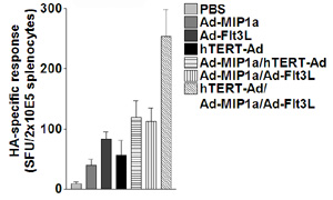
To validate their virotherapy design, the researchers performed IFN-gamma ELISpot experiments using thinkpeptides’ purified peptides covering the HA and OVA epitope sequences. Their results show that infection of tumors with actively replicating virus was an efficient way of generating HA- and OVA-specific IFN-gamma-producing CD8+ T cells. Combining virotherapy and cytokine expression gave rise to an even greater number of epitope-specific CD8+ T cells (figure 1).
|
|
Figure 1: Immune response against intratumoral HA antigen measured by IFN-gamma ELISpot assays after incubation with MHC class I–restricted HA peptide. Data show responses measured from mice treated with cytokine-expressing adenovirus only, with both cytokines in combination, with replicating model-antigen expressing adenovirus alone, and then with the cytokines and replicating virus in combination, as labeled. |
As a further test of the oncolytic capabilities of the CD8+ T cells from their virus-infected and tumor-bearing mice, custom peptides were used in an in vivo CTL assay. DC were introduced at the tumor sites to further enhance responses in these mice. Target cells were labeled ex vivo with the membrane dye CFSE and pulsed with OVA peptide, and then returned to treated mice via i.v. injection. Loss of the CFSE signal indicates that the target cells have been lyzed, demonstrating antigen-specific cytoxic activity (for example data, see figure 2). The best antigen-specific response was found in mice treated with replicating OVA-expressing adenovirus in combination with cytokines.
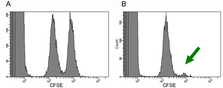
Figure 2. Systemic antitumoral immune response can be measured by CTL assay. Control peptide-pulsed target cells are loaded with a low concentration of CFSE (left peak), for comparison with target peptide pulsed cells, loaded with a higher concentration of CFSE (right peak). (A) results from untreated mice, and (B) results from tumor-bearing, treated mice. Target cells were lyzed in the treated mice, as shown by the disappearance of the ‘target’ peak (green arrow).
In this study Edukulla et al have established a protocol for improving oncolytic virotherapy, using assays for evaluation of tumor response that relied on purified custom peptides from thinkpeptides. A sustained response directed against intratumoral antigens (such as the artificially expressed OVA and HA antigens used in this work) is highly desirable for cancer therapy, and the increased response that this team induced through stimulating DC recruitment and maturation makes this strategy even more attractive. It now remains to see if their approach can be successful in a clinical setting, against ‘real’ antigens.
This work was carried out at the Universities of Hannover, Germany, and Iowa, USA
Figures reproduced by kind permission of Norman Woller (Medizinische Hochschule Hannover)