Your basket is currently empty!
Research into
Research into Type I Diabetes therapy makes use of PEPscreen®technology
|
|
Bresson, D. et al. (2010). Genetic-induced variations in the GAD65 T-cell repertoire governs efficacy of anti-CD3/GAD65 combination therapy in new-onset type I diabetes. Molecular Therapy. 18(2):307-316 [PubMedID: 19690518]
Photo: Dr. Damien Bresson, La Jolla Institute for Allergy and Immunology, USA. |
Bresson et al. investigated a novel combined therapy for type I diabetes consisting of treatment with anti-CD3 antibody plus a DNA vaccination with islet auto-antigen glutamic acid decarboxylase 65 (hGAD65). Mice from a C57BL/6 background and a NOD background were treated with either the combination therapy or individual anti-CD3 or GAD65 DNA vaccination.
GAD65-induced regulatory T cells (Tregs), splenocytes and pancreatic lymph node cells from treated mice underwent an in vitro proliferation assay using a PEPscreen® library of overlapping peptides from the hGAD65 sequence as a stimulant. The peptides had been pooled into 11 groups of 5 or 6 individual peptides. Combination therapy with anti-CD3/GAD65 markedly increased proliferation with 2 of the 11 peptide pools and to a lesser extent with a further 4 peptide pools when compared with individual therapies. The peptide pools that showed the highest response levels included previously described MHC class II immunodominant GAD65 epitopes (p27398-430 and p35524-543). These increased responses were only observed in the mice from a C57BL/6 background, correlating with an increase in GAD65 specific Tregs, whilst no differences between the combination and mono-therapies were observed in mice from a NOD background. This indicates that Treg responses to this combined treatment may be dependent on the MHC complexes expressed in an individual, highlighting the requirement to assess potential therapeutic functionality on a variety of genetic backgrounds.
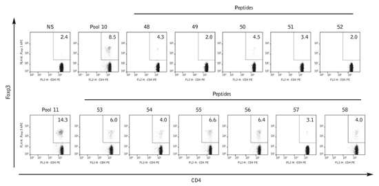
Figure 1:
Glutamic acid decarboxylase of 65 kd (GAD65)-specific regulatory T cells (Tregs) expanded in the RIP-LCMV-GP mice after combination therapy (CT) primarily recognize the C-terminal region of human GAD65 (hGAD65). Splenocytes and pancreatic lymph node cells were derived from NOD- and RIP-LCMV-GP-protected mice 4 weeks after treatment with NM-anti-CD3 alone or in combination with pCMV/hGAD65 (CT). Splenocytes from RIP-LCMV-GP mice protected upon CT were stimulated with two pools of peptides (pools 10 and 11) or the single peptide from the pools 10 and 11 (covering the sequence 471–541 of hGAD65). On day 5 after stimulation, the percentage of Tregs was acquired by flow cytometry. Numbers shown in each histogram correspond to the percentage of Foxp3+ in the CD4+ T-cell population. Data are representative of four independent experiments. GP, glycoprotein; LCMV, lymphocytic choriomeningitis virus; NOD, nonobese diabetic; RIP, rat insulin promoter.
Reprinted by permission from Macmillan Publishers Ltd [Molecular Therapy] Copyright (2010).