
Your basket is currently empty!
Ankyron PECA1_HUMAN Clone AC40115
Clone Information
|
Clone ?
|
AC40115 |
|
Gene
|
PECAM1 |
|
UniProtID
|
P16284 |
|
Organism
|
Homo sapiens (Human) |
|
Entry Name
|
PECA1_HUMAN |
|
SNR ?
|
238 |
|
Product Type
|
Monomer |
|
Ankyron Applications
|
F, IF, ELISA, Magnetic bead sorting |
|
Pack Size
|
100ug |
|
Label ?
Clones are also labelled with a His tag by default
|
Ankyrons® Testing Data
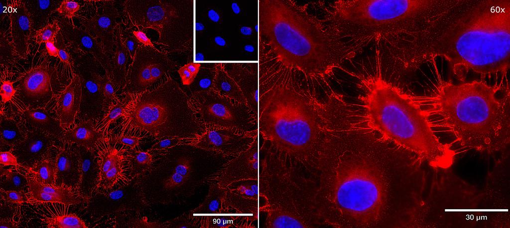
HUVEC were fixed in formaldehyde (4 %), then stained using 1.4 µg V5-tagged Ankyron and anti-V5 antibody conjugated with APC (red) in Staining Buffer (RPMI 1640 + 10 % human serum), counterstaining nuclei with DAPI (blue). Inset: negative control of anti-V5 APC antibody alone.
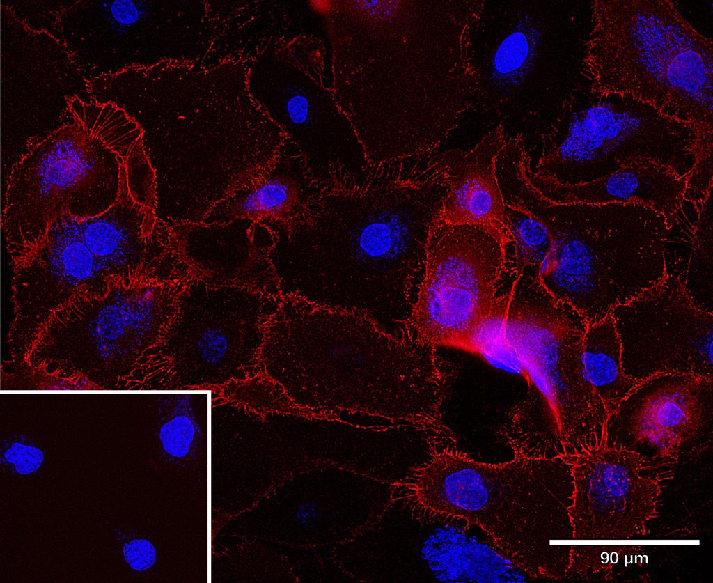
HUVEC were fixed in formaldehyde (1%), then stained using 1.4 µg V5-tagged Ankyron and anti-V5 APC (red) in Staining Buffer (RPMI 1640 + 10 % human serum), counterstaining nuclei with DAPI (blue). Inset: negative control of anti-V5 APC alone.
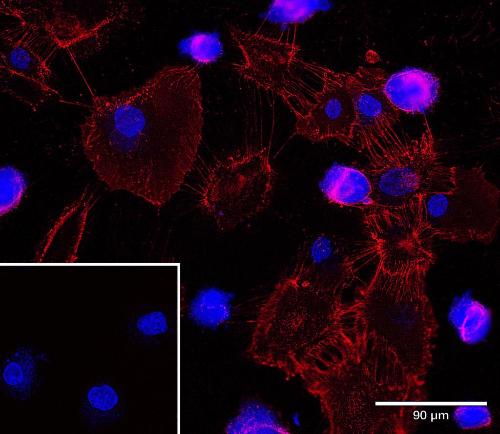
HUVEC were stained using 1.4 µg V5-tagged Ankyron and anti-V5 APC (red) in Staining Buffer (RPMI 1640 + 10 % human serum), counterstaining nuclei with DAPI (blue). Inset: negative control of anti-V5 APC alone.
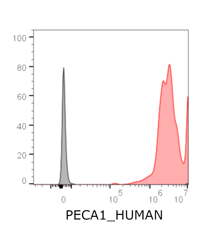
Flow cytometric analysis of HUVEC following staining with pre-conjugated Ankyron (0.4 µg) and anti-V5 APC (red). Negative control, anti-V5 APC only (grey).
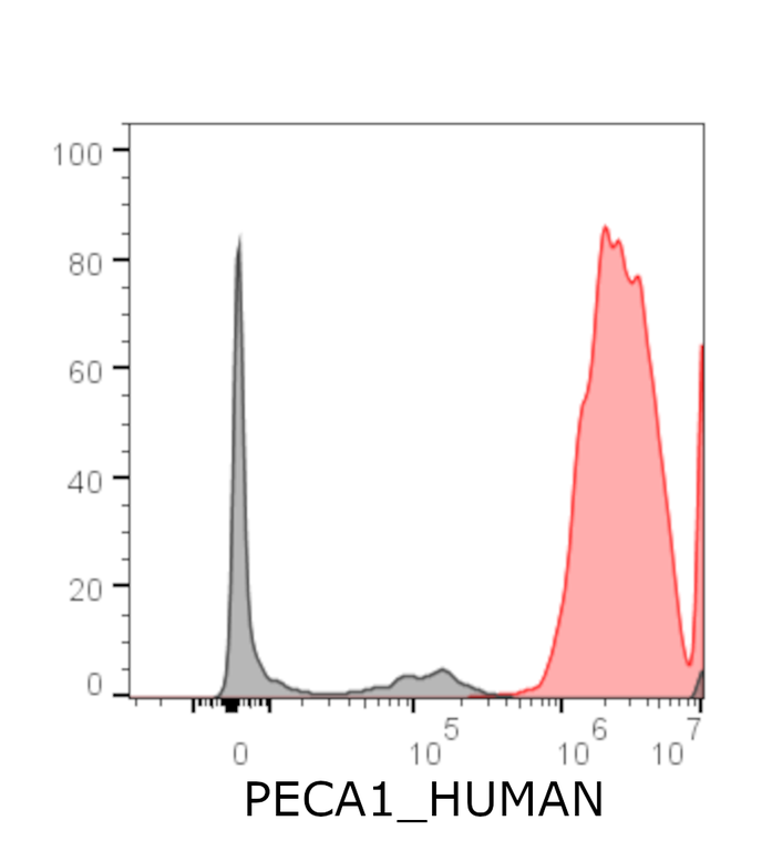
Flow cytometric analysis of HUVEC following fixation in formaldehyde and staining with pre-conjugated Ankyron (0.4 µg) and anti-V5 APC (red). Negative control, anti-V5 APC only (grey).
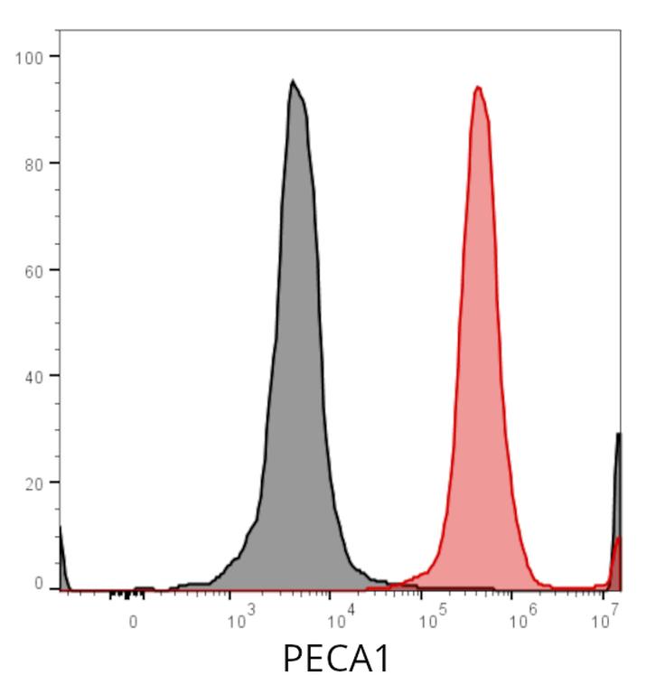
Flow cytometric analysis of HUVECs following fixation with formaldehyde (1%) and subsequent co-incubation of 0.025 μg Ankyron with anti-V5 APC(red). Negative control, anti-V5 APC only.
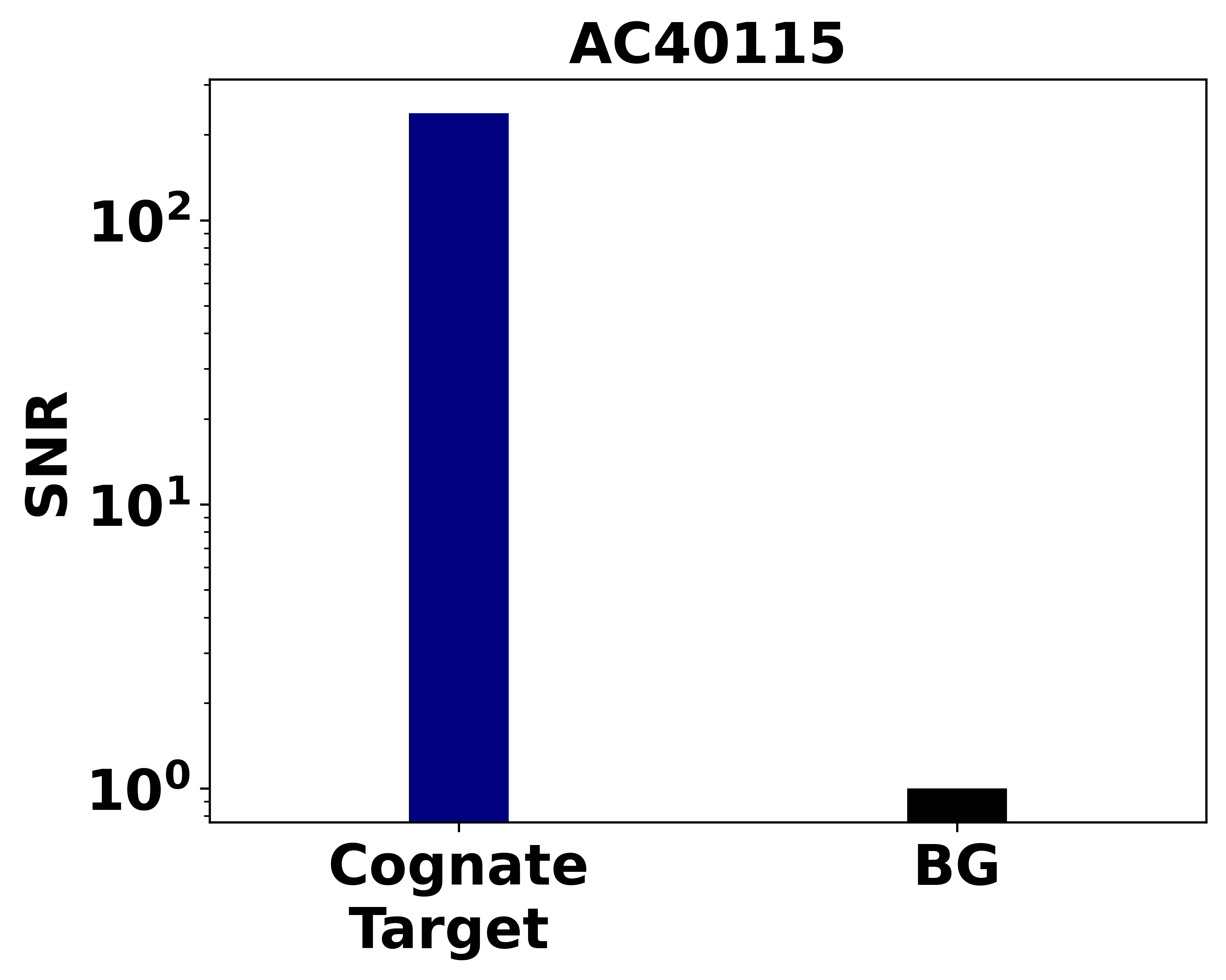
Binding assay determination of the signal to noise ratio (SNR) of V5-labeled Ankyrons® AC40115 binding to 0.5 picomoles of immobilised PECA1_HUMAN, over a background (BG) of non-cognate target protein. The Ankyron was pre-conjugated with the ProImmune Anti-V5 HRP label reagent before detection with a chemiluminescence substrate.
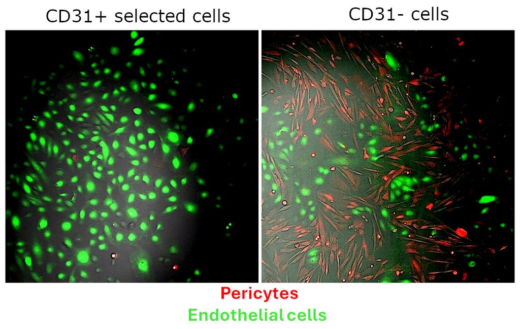
CD31+ endothelial cells were selected by magnetic beads, following labeling with biotinylated Ankyron (2 µg, 15 min) and streptavidin microbeads ( 1 in 10, 15 min) and separation on a magnetic column. Over 96 % purity was achieved (comparable to that obtained following sorting using an antibody), with pericytes (red) visible only in the CD31-negative flow-through cells. Data courtesy of Natalie King and Dr. Paul Holloway.